8.07.2026
Ki 67 – Why a Well-Known Breast Cancer Marker Matters More Than We Thought
Ki-67 is one of the most widely used biomarkers in breast cancer diagnostics. For decades, physicians have relied on it to estimate how actively a tumor is growing. Now, a new study led by Prof. Nicola Aceto and colleagues at ETH Zurich suggests that Ki-67 plays a broader role in breast cancer progression than previously recognized.
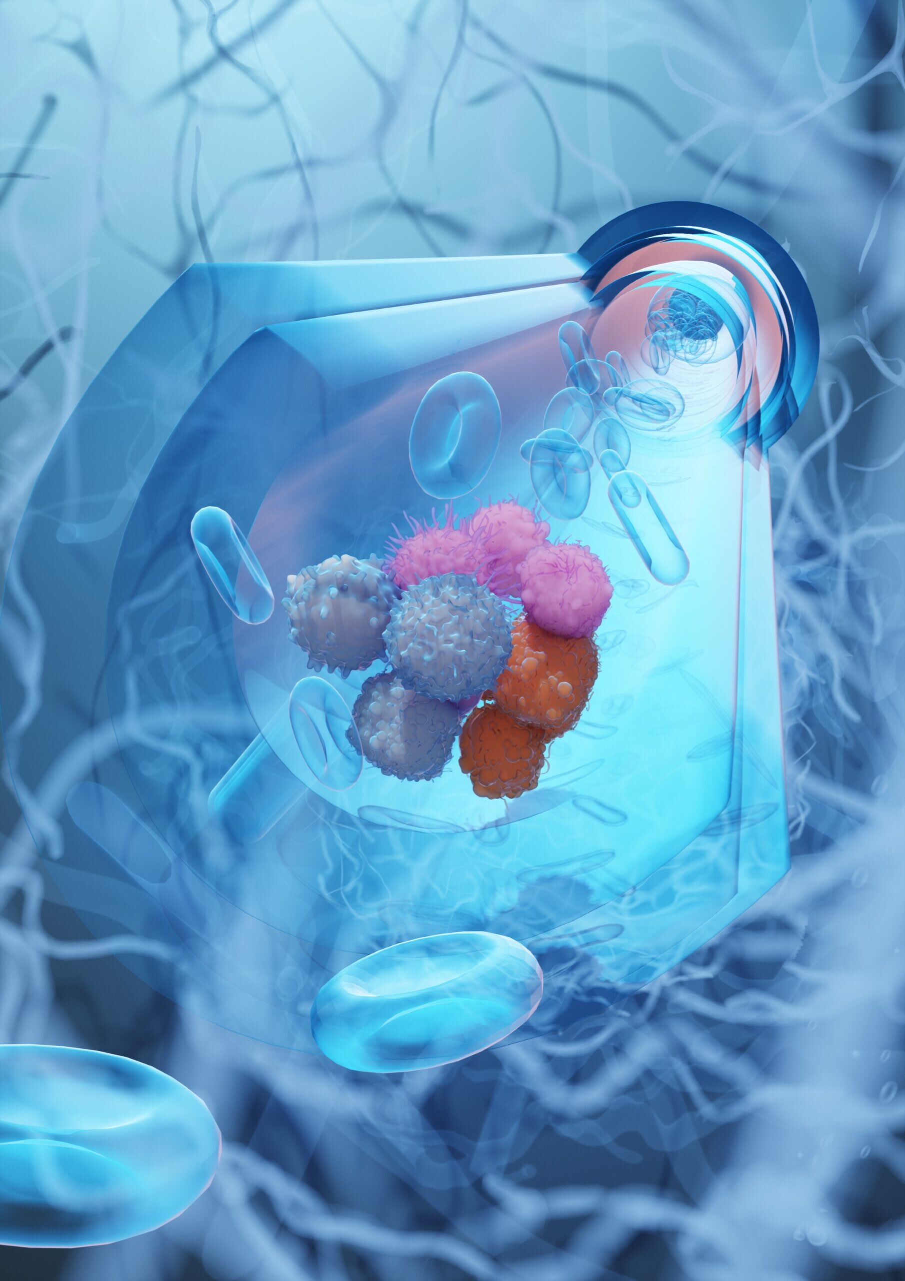
Illustration by Ella Maru Studio
It does not merely indicate how fast a tumor is growing – it promotes the escape of cancer cells into the bloodstream and the formation of metastases. These findings could ultimately help identify the most dangerous cancer cells more precisely and prevent their spread.
What Is Ki‑67 – and What Does It Measure?
In many patients with breast cancer, tissue samples are routinely analyzed for the so-called Ki-67 proliferation index. Ki‑67 is a protein found in the cell nucleus that signals whether a cell is actively dividing. The proportion of tumor cells expressing Ki-67 provides an estimate of how actively the tumor is growing – the more Ki-67, the more rapidly the tumor grows. Higher Ki-67 levels are generally associated with a more aggressive disease course and are used alongside other clinical and pathological factors to guide treatment decisions.
Until now, Ki-67 was viewed primarily as a readout of cell proliferation rather than as influencing cell behavior itself.
Cancer Cells in the Blood
For breast cancer to form metastases, cancer cells must leave the original tumor, penetrate blood vessels, and travel through the bloodstream to other organs. These cells are called circulating tumor cells (CTCs); they can appear as single cells or in small groups known as clusters – and clusters have a particularly high potential to seed new tumors.
In the new study, published in the journal Cell Reports, ETH Zurich researchers analyzed thousands of breast cancer cells and found that Ki‑67 is especially abundant in CTC clusters. This raised an important question: could Ki-67 be helping cancer cells spread?
Switching Off Ki‑67 – and Reducing the Spread
To find out, Yongzhan Zhang, together with colleagues from the Aceto laboratory at ETH Zurich, genetically removed Ki-67 from breast cancer cells in mouse models,
then observed what happened. Surprisingly, the original tumor continued to grow – but far fewer cancer cells entered the bloodstream, and significantly fewer metastases developed. Ki-67 therefore appears to support programs within the cell that make it easier for cancer cells to stick together, invade blood vessels, and drive metastasis.
New Therapeutic Targets: CD47 and KLF4
To better understand how Ki‑67 drives this process, the researchers looked for genes that act downstream of Ki‑67. They identified two particularly important molecules: CD47 and KLF4. CD47 is a surface protein that helps cancer cells adhere to one another, among other roles. KLF4 is a so-called transcription factor that likewise promotes cell-to-cell cohesion. When Ki‑67 is absent, CD47 and KLF4 also become less active – and cancer cells lose the ability to form clusters and invade blood vessels. When CD47 or KLF4 are artificially reactivated in Ki‑67-deficient cells, the capacity to metastasize returns.
Clinical Relevance
Collectively, these findings are clinically significant: while Ki-67 itself may be difficult to target pharmacologically, CD47 is already being investigated as a therapeutic target in early clinical trials. In the long term, therapies that specifically block CD47 or KLF4 could help prevent the spread of breast cancer at an early stage – an approach that aligns well with the goals of the CCCZ: not only to treat cancer, but to stop its spread at the source.